

Morrissey College of Arts & Sciences
Grounded in the liberal arts, the Morrissey College offers undergraduate and graduate programs in the humanities, social sciences, and natural sciences, preparing students to thrive in a broad range of careers while using their gifts to enhance the common good.
Undergraduate Studies at a Glance
37
Majors
48
Minors
22
Academic Departments
4
Special Programs
Liberal Arts Advantage
BC's approach to liberal arts education challenges students to learn across disciplines, prepares them for a range of rewarding careers, and helps them discern who they want to be—and why.
The Core Curriculum
The centerpiece of Jesuit education has always been a common curriculum that emphasizes the rigorous study of the defining works of the humanities, natural sciences, and social sciences.
Graduate Programs at a Glance
25+
Graduate Programs
585+
Doctoral Students
190+
Master's Students
100%
Funding for Doctoral Students
News


Belonging at BC
Our community is made richer by the diversity of our students, faculty, and staff, whose unique perspectives and lived experiences contribute to the vibrancy of our campus and classrooms.
Discover Boston College


Students reflect during a retreat at the Connors Center
Bapst Library has been nationally recognized as one of the most beautiful libraries in higher education
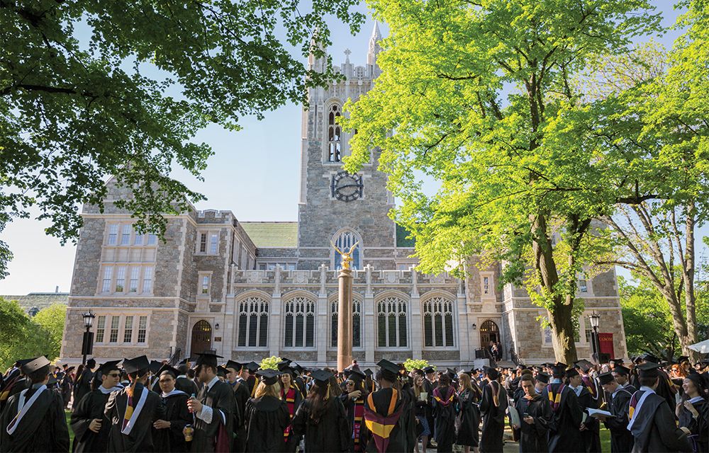
Students prepare for graduation on Linden Lane
Fr. Kalscheur meeting with students in Gasson Hall.
Professor Ethan Baxter in the lab with students.
The PULSE program educates students about social injustice by putting them into direct contact with marginalized populations.
Academic convocation is a Boston College tradition celebrating the start of the academic year.
Leading the Way
Boston College is at the forefront of scientific research and innovation, while also prioritizing a formative liberal arts education as the foundation for our R1 Research acumen. Across disciplines, collaborative teams of students and faculty are examining the complex problems of our contemporary world, proposing new solutions and new ways of thinking about religion and culture, science and technology, art and education, business and ethics.
Giving
To make a donation to the Morrissey College of Arts and Sciences, please visit the University's Giving website.